loader image
Poppy is a four-year-old female spayed Pitt Bull Mix. She was quilled by a porcupine on the right side of her face in late December 2025. A few weeks after the quilling, she developed some jaw pain. She had a head CT, which showed no major changes, but a few more quills were found on an explore. A few weeks after that (now about six weeks after the quill incident), Poppy developed seizures and her owner brought her to MVMC. On exam, Poppy clearly had an issue going on with her right forebrain. We performed an MRI, which showed an collection of abscesses in her ventral right forebrain, with a small skull defect leading to them. We assumed that, somehow, a quill had bored its way through Poppy’s skull even though this is not typical quill behavior to go through bone (they like the path of least resistance).

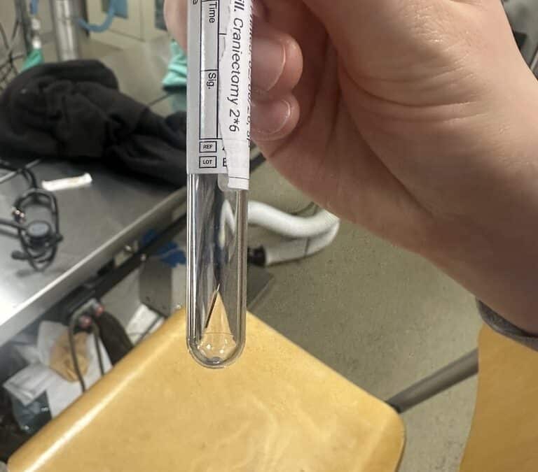
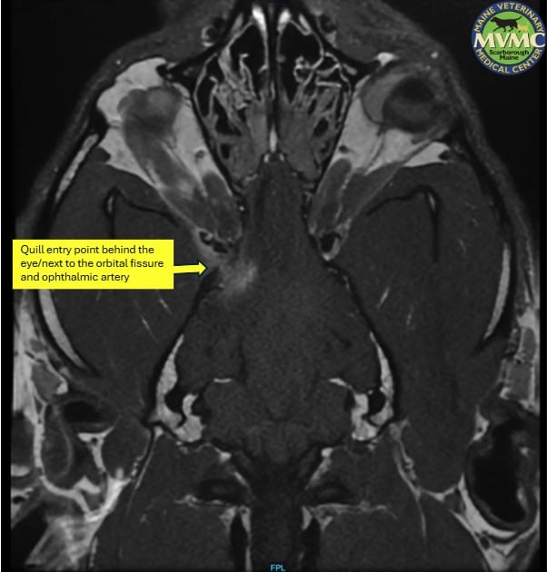
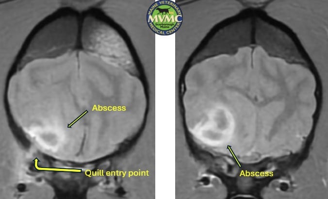
Quills are not visible on MRI or any imaging modality besides occasionally ultrasound. Unfortunately, the location of the abscess/skull penetration was very difficult to access directly – it was right behind Poppy’s eye socket, about a millimeter behind a hole in the skull where important nerves and blood vessels enter/exit called the orbital fissure.

We initially tried Poppy on medical management (antibiotics and steroids), but her neurological status showed signs of deterioration, so we repeated an MRI two days later and the abscesses had grown in size and her brain tissue was being shifted in a dangerous way by the mass effect.

CT Scan
We made the decision to make a craniectomy in the top of Poppy’s skull on the right side at the level of the abscesses and use Dr. Lemon’s ultrasound expertise to visualize the abscesses through Poppy’s brain tissue. The initial goal of the surgery was to use a needle to perform an ultrasound-guided abscess drainage/obtain a culture sample and flush the infected tissue gently. When Dr. Lemon had the ultrasound on the abscesses, we could see a seven-millimeter tubular structure straddling between each abscess and were astounded that it looked like we could visualize the culprit quill.
2 CT-Scan
We first drained the abscesses successfully, but I felt we had to make an effort to remove the quill to mitigate the risk of recurrent infection/further quill migration causing brain injury. Using a long-necked biopsy instrument with a grabber on the end, Dr. Lemon was able to guide the instrument to the quill so that we could pull it out!

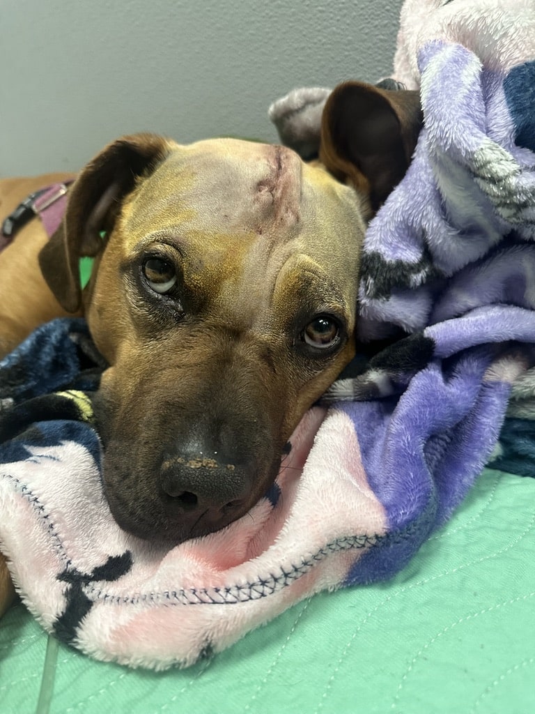
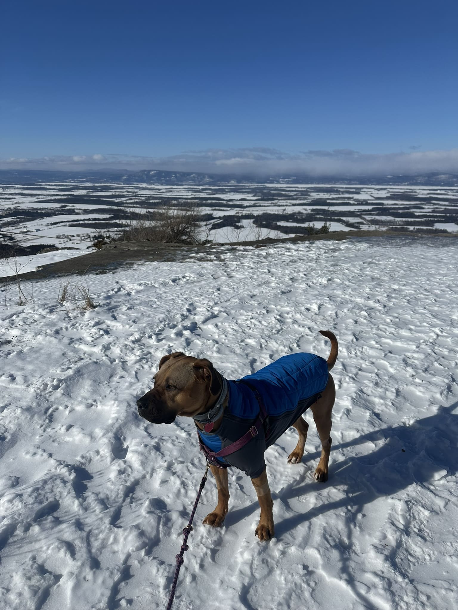
Poppy did great after surgery, although she did have some transient blindness on her left side due to some inflammation in her thalamus on the right side caused by the surgery. That resolved within about a week and Poppy now had a normal neurological status.

We performed a repeat MRI a couple weeks ago, and you’d almost never know she had this terrible problem besides for an obvious skull defect from the surgery, and a very faint tract of blood product leftover from the path of the biopsy instrument/needle used to drain the abscess. Her healing has been really remarkable.

I’m incredibly proud of this case because: a) this type of intervention has never been described/probably never happened before, b) it took the whole team to make it happen/coordination between several departments including both doctors and nurses, and c) the outcome has exceeded anyone’s expectation given how difficult this presentation was.

dog partially covered with a blanket
The fact that it was a long-time friend’s dog is the icing on the cake. She has been incredibly dedicated to Poppy’s follow-up care (i.e. all the re-imaging — Poppy has had three MRIs now and we plan on a fourth in about six weeks). I have shared this case with my neuro colleagues around the country and they all agree that my career has probably peaked/maybe I should just retire 😊. – Scott Petesch, VMD, DACVIM (Neurology)
a dog standing on the sand